Written by

Stem cells age. But not in the way we think.
As we get older, the body’s regenerative capacity falls behind. It’s easy to assume our stem cells simply run out, that the repair crew dwindles until there’s no one left to rebuild.
The truth is more complicated.
In the bone marrow, hematopoietic stem cells (HSCs), the source of every blood and immune cell, don’t vanish with age. They multiply [1].
In mice, the number of HSCs can increase ~9-fold with age, while each cell’s regenerative output drops roughly 3-fold [2]. In other words, far more cells, but less productive ones.†
That paradox led scientists to an experiment straight out of a gothic novel.
In 2005, researchers at Stanford stitched young and old mice together so that they shared a single bloodstream, a setup called heterochronic parabiosis [3]. For five weeks, two animals lived as one, their systems intertwined.
When the older mouse’s muscles were injured, they healed almost as fast as the young one’s, forming new fibers twice as effectively as before. The liver, too, ramped up cell renewal 2- to 3-fold. But the rejuvenation wasn’t from young cells migrating in. Less than 0.1% of the new tissue carried any trace of the young partner. The old cells were doing the work themselves.
The finding was truly profound: aging stem cells hadn’t lost their potential, only their instructions (stem cell exhaustion). When those molecular messages were restored, regeneration returned.
The lesson here wasn’t that youth can be transfused. It was that signaling can be renewed.
The bloodstream carries an operating code that tells stem cells when to rest, divide, and repair. Change that code, and you change the body’s capacity to heal. And those same molecular signals, which were manipulated artificially via parabiosis, can also be modulated naturally through how we live: how we sleep, how we move, and what we eat.
In this article, we’ll explore how everyday behaviors influence that regenerative code:
how sleep synchronizes stem cell timing,
how movement mobilizes them into circulation,
and how nutrition shapes the signals that decide whether we heal or stagnate.
Sleep and Stem Cells: How Rest Resets the Body’s Repair Clock
Your stem cells keep time.
So does nearly every other cell in your body, each one following an internal clock. And that circadian rhythm plays a profound role in healing.
When researchers tracked thousands of burn patients, a surprising pattern emerged: wounds that happened at night took 11 days longer to close than those sustained during the day [4]. That staggering difference traced back to fibroblasts — the skin’s weavers of new tissue — whose engines run fastest in daylight, anticipating the damage we’re most likely to incur while awake and active.
The same rhythm extends deep in the bone marrow.
Here, hematopoietic stem cells live under a molecular handshake: CXCR4 receptors on stem cells bind to matching CXCL12 signals from their support cells. The message transmitted here is simple: stay home.
But every morning, a pulse of norepinephrine quiets CXCL12, loosening the bond [5]. Stem cells slip free, enter circulation, and start their daytime patrol. Then when night falls, melatonin from the pineal gland restores CXCL12 expression and that “stay home" signal [6]. The stem cells retreat to their niches, shielded from inflammation, recharging for the next day’s work.
It’s a perfect rhythm: mobilize, rest, repeat. Until sleep falls short.
In one experiment, losing four hours of sleep was enough to weaken stem cells at their core [7]. When transplanted into mice whose marrow had been wiped out, these sleep-deprived stem cells rebuilt only about half as much of the new blood system as normal.
But it’s not just total sleep time that matters. Consistency is critical as well.
When sleep is fragmented, the body’s clocks fall out of sync. The brain keeps time, but the body stops listening, and those signals that tell stem cells when to stay put and when to mobilize turn noisy and confused.
In mice, fragmented sleep caused bone marrow to lose its regenerative cadence. Stem cells that should have cycled between rest and renewal stayed half-awake, working when they should have been recovering. Within weeks, the marrow started making more myeloid cells — immune cells that drive inflammation — and fewer of the stem cells that sustain long-term repair [8]. The blood’s balance tilted toward the pattern we typically see in aging.
Worse yet, the change persisted. Even after three months of normal sleep, the marrow hadn’t fully reset. When those stem cells were transplanted into healthy mice, they churned out the same inflammatory profile. The change ran deep: chemical marks on their DNA had rewired how they read their genes, leaving behind a lasting cellular memory of sleeplessness.
That’s why timing and quality matter as much as hours. A solid block of restorative sleep resets the system each night: melatonin rises, the “stay home” signal strengthens, and stem cells repair themselves before the next day’s work.
By morning, those clocks strike together again. Stem cells are back on schedule, blood flow quickens, and the body’s renewal machinery shifts from rest to activity.
Aerobic Exercise and Stem Cells: How Movement Keeps Blood Vessels Young
If sleep restores the body’s rhythm, movement carries that rhythm outward.
With each heartbeat, blood delivers oxygen, but more than that it carries signals. The force of its flow across vessel walls keeps those walls responsive to their environment, adjusting to pressure and chemistry.
Blood vessels, after all, are not plumbing. They’re living tissue, constantly listening to flow. With every beat, the blood’s movement brushes against the inner lining — the endothelium — creating what physiologists call shear stress: a kind of good friction that keeps the lining alert and engaged.
That sensitivity, however, dulls with age, and sooner than you might think. Endothelium-dependent vasodilation begins to decline in our twenties [9], and inactivity accelerates the slide [10].
The arteries of sedentary men in their fifties and seventies were found to dilate about 25% less than peers in their twenties. But in men who exercised regularly, that age gap vanished. Their blood vessels reacted as if time had stood still.
Even more striking: after just three months of moderate aerobic exercise, previously sedentary men fully regained that responsiveness. Vasodilation rose by roughly 30%, restoring endothelial function to youthful levels [11].
The question is — how?
Beneath the surface, aerobic training reawakens the body’s repair economy. It stirs endothelial progenitor cells (EPCs), stem-like builders that patch vessel walls and help new ones grow.
In sedentary adults, those cells tend to be scarce and sluggish. Their colonies shrink by about 75%, and their ability to migrate toward injury drops by nearly half. But after just a few months of steady aerobic exercise, EPC formation doubles, and mobility improves by roughly 50% [12].
Animal models reveal how this works. In mice with access to running wheels, circulating endothelial progenitor cells tripled within weeks. But when scientists blocked nitric oxide signaling, the effect vanished [13].
That’s the key. Shear stress from blood flow activates nitric oxide, which relaxes the vessels, increases flow, and signals for repair. Hot on its heels comes VEGF, one of the body’s most potent “come help” molecules. Within hours of exercise, VEGF levels surge, freeing stem-like cells from the bone marrow and sending them into circulation [14].
Through this loop of flow, signaling, and renewal, aerobic exercise keeps the vasculature — and the body’s regenerative dialogue — alive.
HIIT and Stem Cells: How Intensity Primes the Body to Heal Faster
Steady movement lays the groundwork. Intensity teaches the system to respond.
Brief surges of near-maximal effort, like those in high-intensity interval training (HIIT) — send the body’s repair network into overdrive, a stress rehearsal that sharpens its reflexes.
Researchers tested this by comparing two cycling sessions matched for total work: ~45 minutes of steady pedaling versus ~15 minutes of much harder intervals. The long ride barely moved the needle. In contrast, the short intense one triggered a surge of stem cells from the bone marrow, roughly 2.5 times the usual number in circulation [15]. Intensity, not duration, flipped the switch.
The mechanism traces back to the body’s stress chemistry. The same norepinephrine that wakes the marrow each morning floods the system during hard effort, carried on waves of adrenaline that raise pulse and pressure [16]. The marrow hears that signal and releases repair cells into circulation — a coordinated response between the nervous system and the body’s regenerative core.
Beyond mobilizing stem cells, HIIT may also prolong the window in which these repair cells can do their work.
In one experiment, athletes completed a classic HIIT workout: four rounds of high-intensity intervals, the infamous 4x4 protocol [17]. Within minutes, repair cells flooded the bloodstream, while levels of CD31, a protein that normally helps those cells return to the marrow, dropped by about 10%. With that exit signal muted, the cells lingered longer, patrolling the bloodstream for damage before heading home.
Over time, these bouts of brief stress lead to regenerative reprogramming.
Trained athletes, conditioned by years of high-intensity work, have been shown to carry 3- to 4-fold higher baseline repair cells than sedentary peers [18]. Their marrow, in effect, has learned the rhythm of exertion, keeping a deeper pool of regenerative cells on standby.
Mediterranean Diet and Vascular Repair: How Food Fuels Regeneration
Exercise primes the system to rebuild. Diet decides what it rebuilds with.
Aging blood vessels reflect a tug-of-war between damage and repair. The endothelium continually renews itself, shedding fragments of damaged tissue and replacing them with new cells. The debris shows up in the blood as endothelial microparticles, tiny shards of damaged tissue. The repair crew arrives as endothelial progenitor cells, sent to patch and rebuild. When injury outpaces repair, the vessels stiffen, and blood flow begins to fail.
Could the foods we eat shift the balance?
In a crossover trial, older adults rotated through three diets: four weeks of a Mediterranean pattern rich in virgin olive oil, four weeks high in saturated fat from butter, and four weeks low-fat with added plant omega-3s [19].
Only the Mediterranean diet changed the equation, tilting it toward repair.
Endothelial progenitor cells roughly doubled while vascular debris fell by about half. Blood flow responses improved and oxidative stress markers dropped. Levels of the antioxidant enzyme superoxide dismutase (SOD) also declined — not from depletion, but because there was less oxidative chaos to defend against.
And this boost in repair capacity may be linked to better vascular structure over time.
In a year-long follow-up, people who stuck with the Mediterranean pattern kept those repair signals active, and the structure of their blood vessels began to show it. The walls of the carotid artery, a tissue that often thickens with age, shifted in the opposite direction, hinting that steady dietary cues can help the vasculature preserve its youthfulness [20].
So what, exactly, gives the Mediterranean pattern its restorative power?
Healthy Fats and Stem Cells: How Olive Oil Supports Vascular Renewal
Olive oil sits at the heart of the Mediterranean diet.
Its benefits are often credited to the peppery phenolics that give extra-virgin oil its bite [21]. But even stripped of antioxidants, olive oil’s chemistry carries regenerative weight.
In one crossover trial, participants consumed virgin olive oil for four weeks [22]. Their blood vessels responded as if resurfaced: fewer microparticles from stressed vessels, more repair cells in circulation, and stronger blood flow responses. The oil’s antioxidant phenolics were thought to be the drivers of these benefits.
But even when those phenolics are removed, the repair signal persists.
In another study, replacing just 10% of dietary calories from saturated fat with refined olive oil — rich in monounsaturated fat but low in antioxidants — increased progenitor cell counts by nearly 30% and cut vessel-wall debris by half. When the same exchange was made with other unsaturated fats, the repair signal disappeared [23]. Only olive oil produced less damage and more regeneration.
Why? Chemistry.
Olive oil’s dominant fatty acid, oleic acid, carries a bend in its carbon chain that prevents membranes from packing too tightly [24]. That small kink makes cell surfaces more flexible and responsive to nitric oxide, the same signal that mobilizes stem cells from marrow. The result is a more adaptive endothelium, one that resists injury while sustaining the repair dialogue.
Polyphenols and Stem Cells: How Plant Compounds Signal Repair
For decades, the “French paradox” had taunted nutrition scientists. How could populations who buttered everything still boast supple arteries? The answer, some suspected, was hiding in the wine glass.
Sure enough, when researchers asked volunteers to drink a glass of red wine every day — a study that probably didn’t struggle for participants — their blood began to change [25].
In just three weeks, the population of endothelial progenitor cells more than doubled. But more than just multiplying their numbers, they became more functional and resilient. Under the microscope, those same cells moved more freely, signaled more nitric oxide, and were less prone to stress-induced cell death.
But was it the alcohol, or something else?
Researchers compared red wine, beer, and vodka. Same alcohol, same calories. Only the red wine group showed the signature of repair: more circulating endothelial progenitor cells and stronger nitric oxide activity [26].
What separated red wine from the rest wasn’t the alcohol, but the polyphenols, compounds that plants generate to defend and repair under stress. When scientists isolated resveratrol, one of wine’s polyphenols, the story replayed in vitro: EPCs multiplied, migrated, and even began weaving primitive vessel-like networks inside the petri dish [27].
But the vascular benefits of polyphenols aren’t confined to wine or even grapes.
In a double-blind trial, volunteers drank cocoa beverages every day that were identical in every respect except one: polyphenol content [28]. After four weeks, the high-polyphenol version increased vascular responsiveness by nearly 50% and more than doubled the number of circulating repair cells. The scale of that increase matched what’s been seen after major lifestyle changes like exercise or smoking cessation.
Across studies, the pattern holds: polyphenols amplify nitric oxide signaling and sustain the circulation of endothelial repair cells [29].
Together, plant polyphenols and olive oil form a regenerative partnership. Polyphenols enhance the signals for repair, while monounsaturated fats provide the structure for them to take hold.
Rewriting the Regenerative Code
The parabiosis experiments showed us that aging stem cells are not broken. They are misinformed.
We may not have young blood on tap (yet), but many of the same signals restored by parabiosis can be tuned through how we live.
Exercise sends a wave of messages that echo the effects of young blood, boosting nitric oxide and vascular growth cues that help stem cells leave the marrow and rebuild tissue [30].
Sleep restores the body’s timing system, the daily rhythm that tells stem cells when to rest and when to mobilize. It also quiets the inflammatory messengers that accumulate with age [31], the very signals young blood was shown to suppress [32].
Nutrition fine-tunes the environment those cells live in. Diets rich in plant compounds and healthy fats calm the background noise of oxidative stress, recreating some of the same internal chemistry that youthful blood re-established [33]. Once those foundations are in place, adding targeted nutraceutical support can help you boost repair with stem cell pathways that are already being activated by sleep, movement, and diet.*
If you want to start tuning your own regenerative signals:
Keep your rhythms steady. Get morning light, dim screens before bed, and go to sleep and wake up at consistent times.
Move most days. Aim for about 150 minutes of moderate activity each week to build your aerobic base, plus 1–2 short high-intensity sessions.
Eat like the Mediterranean. Use olive oil as your main fat, include nuts and fish, and make plants your foundation.
Feed your vessels. Get polyphenols daily, like from berries, pomegranate, dark chocolate, or green tea.
Support your cellular environment. Beyond diet, monthly targeted stem cell supplements can help maintain the signals stem cells rely on for repair and renewal.*
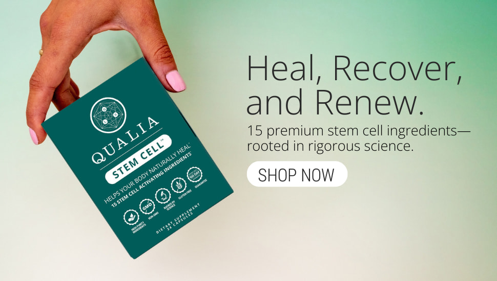
For those who want to take that foundation further, Qualia Stem Cell was developed around the same insight that emerged from parabiosis: our cells’ repair potential depends on the environment they inhabit.*
Qualia Stem Cell was created to help revive your body’s own repair crew: the stem cells and signaling chemistry that keep renewal possible.*
Formulated to support the major pillars of stem cell vitality — survival, renewal, mobilization, and differentiation — through a carefully calibrated blend of fifteen premium ingredients.*
We can’t transfuse youth. But we can cultivate the chemistry that reminds our cells how to repair.*
† Not all stem cell populations follow this pattern. Others actually do decline in number as well as regenerative capacity. Endothelial progenitor cells, for instance, can fall by as much as 70–90% from youth to old age.
*These statements have not been evaluated by the Food and Drug Administration. This product is not intended to diagnose, treat, cure, or prevent any disease.
References
[1] B. Chatterjee, S.S. Thakur, in: S. Pathak, A. Banerjee (Eds.), Stem Cells and Aging, Academic Press (2021) 103–111.
[2] S.M. Chambers, C.A. Shaw, C. Gatza, C.J. Fisk, L.A. Donehower, M.A. Goodell, PLoS Biol. 5 (2007) e201.
[3] I.M. Conboy, M.J. Conboy, A.J. Wagers, E.R. Girma, I.L. Weissman, T.A. Rando, Nature 433 (2005) 760–764.
[4] N.P. Hoyle, E. Seinkmane, M. Putker, K.A. Feeney, T.P. Krogager, J.E. Chesham, L.K. Bray, J.M. Thomas, K. Dunn, J. Blaikley, J.S. O'Neill, Sci. Transl. Med. 9 (2017) eaal2774.
[5] C.M. Hoffman, L.M. Calvi, Mol. Endocrinol. 28 (2014) 1592–1601.
[6] S. Méndez-Ferrer, D. Lucas, M. Battista, P.S. Frenette, Nature 452 (2008) 442–447.
[7] A. Rolls, W. Pang, I. Ibarra, D. Colas, P. Bonnavion, H.C. Heller, I.L. Weissman, L. de Lecea, Brain Behav. Immun. 32 (2013) e6.
[8] C.S. McAlpine, M.G. Kiss, F.M. Zuraikat, D. Cheek, G. Schiroli, H. Amatullah, P. Huynh, M.Z. Bhatti, L.P. Wong, A.G. Yates, W.C. Poller, J.E. Mindur, C.T. Chan, H. Janssen, J. Downey, S. Singh, R.I. Sadreyev, M. Nahrendorf, K.L. Jeffrey, D.T. Scadden, K. Naxerova, M.P. St-Onge, F.K. Swirski, J. Exp. Med. 219 (2022) e20220081.
[9] S. Taddei, A. Virdis, P. Mattei, L. Ghiadoni, C.B. Fasolo, I. Sudano, A. Salvetti, Hypertension 29 (1997) 736–743.
[10] G. Guhanarayan, J. Jablonski, S. Witkowski, J. Appl. Physiol. 117 (2014) 500–506.
[11] C.A. DeSouza, L.F. Shapiro, C.M. Clevenger, F.A. Dinenno, K.D. Monahan, H. Tanaka, D.R. Seals, Circulation 102 (2000) 1351–1357.
[12] G.L. Hoetzer, G.P. Van Guilder, H.M. Irmiger, R.S. Keith, B.L. Stauffer, C.A. DeSouza, J. Appl. Physiol. 102 (2007) 847–852.
[13] U. Laufs, N. Werner, A. Link, M. Endres, S. Wassmann, K. Jürgens, E. Miche, M. Böhm, G. Nickenig, Circulation 109 (2004) 220–226.
[14] T.P. Gavin, C.B. Robinson, R.C. Yeager, J.A. England, L.W. Nifong, R.C. Hickner, J. Appl. Physiol. 96 (2004) 19–24.
[15] J.M. Baker, J.P. Nederveen, G. Parise, J. Appl. Physiol. 122 (2017) 182–190.
[16] N.H. Agha, F.L. Baker, H.E. Kunz, R. Graff, R. Azadan, C. Dolan, M.S. Laughlin, C. Hosing, M.M. Markofski, R.A. Bond, C.M. Bollard, R.J. Simpson, Brain Behav. Immun. 68 (2018) 66–75.
[17] J.M. Kröpfl, F.G. Beltrami, H.J. Gruber, I. Stelzer, C.M. Spengler, Front. Physiol. 11 (2020) 308.
[18] M.R. Bonsignore, G. Morici, A. Santoro, M. Pagano, L. Cascio, A. Bonanno, P. Abate, F. Mirabella, M. Profita, G. Insalaco, M. Gioia, A.M. Vignola, I. Majolino, U. Testa, J.C. Hogg, J. Appl. Physiol. 93 (2002) 1691–1697.
[19] C. Marin, R. Ramirez, J. Delgado-Lista, E.M. Yubero-Serrano, P. Perez-Martinez, J. Carracedo, A. Garcia-Rios, F. Rodriguez, F.M. Gutierrez-Mariscal, P. Gomez, F. Perez-Jimenez, J. Lopez-Miranda, Am. J. Clin. Nutr. 93 (2011) 267–274.
[20] M.I. Maiorino, G. Bellastella, M. Petrizzo, M. Gicchino, M. Caputo, D. Giugliano, K. Esposito, Eur. J. Prev. Cardiol. 24 (2017) 399–408.
[21] K.L. Tuck, P.J. Hayball, J. Nutr. Biochem. 13 (2002) 636–644.
[22] K. Sarapis, E.S. George, W. Marx, H.L. Mayr, J. Willcox, T. Esmaili, K.L. Powell, O.S. Folasire, A.E. Lohning, M. Garg, C.J. Thomas, C. Itsiopoulos, G. Moschonis, Eur. J. Nutr. 61 (2022) 1073–1086.
[23] M. Weech, H. Altowaijri, J. Mayneris-Perxachs, K. Vafeiadou, J. Madden, S. Todd, K.G. Jackson, J.A. Lovegrove, P. Yaqoob, Am. J. Clin. Nutr. 107 (2018) 876–882.
[24] C. Santa-María, S. López-Enríquez, S. Montserrat-de la Paz, I. Geniz, M.E. Reyes-Quiroz, M. Moreno, F. Palomares, F. Sobrino, G. Alba, Nutrients 15 (2023) 224.
[25] S. Hamed, J. Alshiek, A. Aharon, B. Brenner, A. Roguin, Am. J. Clin. Nutr. 92 (2010) 161–169.
[26] P.H. Huang, Y.H. Chen, H.Y. Tsai, J.S. Chen, T.C. Wu, F.Y. Lin, M. Sata, J.W. Chen, S.J. Lin, Arterioscler. Thromb. Vasc. Biol. 30 (2010) 869–877.
[27] X.B. Wang, J. Huang, J.G. Zou, E.B. Su, Q.J. Shan, Z.J. Yang, K.J. Cao, Clin. Exp. Pharmacol. Physiol. 34 (2007) 1109–1115.
[28] C. Heiss, S. Jahn, M. Taylor, W.M. Real, F.S. Angeli, M.L. Wong, N. Amabile, M. Prasad, T. Rassaf, J.I. Ottaviani, S. Mihardja, C.L. Keen, M.L. Springer, A. Boyle, W. Grossman, S.A. Glantz, H. Schroeter, Y. Yeghiazarians, J. Am. Coll. Cardiol. 56 (2010) 218–224.
[29] J.A. Vita, Am. J. Clin. Nutr. 81 (2005) 292S–297S.
[30] L. Katsimpardi, N.K. Litterman, P.A. Schein, C.M. Miller, F.S. Loffredo, G.R. Wojtkiewicz, J.W. Chen, R.T. Lee, A.J. Wagers, L.L. Rubin, Science 344 (2014) 630–634.
[31] M.R. Irwin, M.R. Opp, Neuropsychopharmacology 42 (2017) 129–155.
[32] E.J. Morrison, D.P. Champagne, M. Dzieciatkowska, T. Nemkov, J.C. Zimring, K.C. Hansen, F. Guan, D.M. Huffman, L. Santambrogio, A. D'Alessandro, Nutrients 11 (2019) 1337.
[33] J. Rebo, M. Mehdipour, R. Gathwala, K. Causey, Y. Liu, M.J. Conboy, I.M. Conboy, Nat. Commun. 7 (2016) 13363.